The composition of bone, the structure of the long bone and the function of the skeleton
Composition of bone
“Bone itself consists mainly of collagen fibres and an inorganic bone mineral in the form of small crystals” (University of Cambridge 2005).
Bones are living tissue which is made up of connective tissue. The bone tissue is made up of several types of bone cells such as osteoblasts and osteoclasts. Osteoblasts are needed to aid the process of healing and osteoclasts are needed to break down old bone to make room for new bone. Bones contain phosphorus and calcium which are inorganic salts; these salts give the bone strength. The bone also has collagenous fibers to give the bone flexibility.
Get Help With Your Essay
If you need assistance with writing your essay, our professional essay writing service is here to help!
Structure of the long bone
The long bone consists of hyaline cartilage which covers the ends of the bone and stops them rubbing together as well as absorbing shock. The head of the long bone is called epiphysis. Compact bone is hard, dense bone and is the outer layer of the long bone, this gives the hallow part of the bone strength. Cancellous bone is the spongey bone in the long bone, which stores the red bone marrow and this is where blood cells are made. The yellow bone marrow is stored in the marrow cavity of the long bone and this is where white blood cells are made. Cancellous bone looks like honey comb as it is very porous and is easily recognised. The epiphyseal plate is where the bones grow in length. The shaft in the long bone itself is called the diaphysis. The periosteum is a protective layer on the long bone that has no cartilage; this is where the tendons and ligaments connect to (Curran 2016). Below is a picture of the structure of the long bone (LinkedIn Corporation 2017).

Functions of the skeleton
The functions of the skeleton include protection, movement, formation of blood cells and storage.
Protection: Vital organs such as the heart and lungs are protected by the skeleton, the heart and lungs are protected by the sternum (breast bone) and the enclosure of the rib cage. The brain is also protected by the cranium (skull).
Movement: Bones and muscles work together to produce body movement as a lever. Bones and muscles form joints which are needed to move.
Formation of blood cells: This process is called haematopoiesis where blood cells are produced in the bone marrow of some bones.
Storage: Bones store minerals such as calcium and phosphorus which are inorganic salts needed for the strength of bones. These materials are stored in the bones when there is too much present in the blood (Curran 2016).
Types of joints and their functions
There are three different types of joints; Fibrous joints, cartilaginous joints and synovial joints. Their main function is movement of the limbs and stability for example the stability found in the bones of the skull (Boundless 2016).
Fibrous Joints
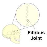
Fibrous joints are immoveable i.e. they cannot move. These joints are held together with only a ligament. An example of this type of joint would be the cranium.
 (Google Images 2017)
Cartilaginous Joints
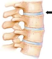
Cartilaginous joints are partly moveable and the connection between articulating bones is made up of cartilage. An example of this type of joint is between the vertebrae that form the spine. The arrow in the below picture indicates where the cartilage is in the spine. (Google Images 2017)
Synovial Joints
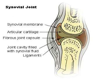
Synovial joints are freely moveable and are the most known and common type of joints in the body. These joints have a synovial capsule surrounding the joint and the synovial membrane secretes synovial fluid to provide lubrication to the joint which prevents friction allows for more movement. Cartilage provides padding to each end of the bone. There are six types of synovial joints; hinge, pivot, ball & socket, saddle, condyloid and gliding joints. Examples of synovial joints would be elbows, knees and hips (Boundless 2016).
 (Google Images 2017)
Cite This Work
To export a reference to this article please select a referencing style below:


