Morphological and Anatomical study of the brain of the african ostrich that between the ages of 7 and 15 month in the city of zabol have been slaughtered
- Dahmardeh Moslem, Akbari Mohammad Ebrahim
Abstract: The morphological and anatomical specifications of the African ostrich brain that between the ages of 7 and 15 month in the city of zabol have been slaughtered were researched in this study. The average weight, length, and width of the total brain are 29.37 gr, 54.30 mm, and 48.09 mm, respectively. The ostrich brain has many more transverse fissures of the cerebellar vermis than do the brains of other poultries. Therefore, the surface area of the African ostrich’s cerebellum is greater. The cerebellum become visible relatively well developed and clearly protrudes dorsally. The posterior superior part of the cerebellar vermis approximately forms an angle of 150°. The cerebrum surface is flat, without any gyrus or sulcus. The gray matter is thin. There is an arcuated telencephalic vallecula on the dorsal surface, and the sagittal eminence is elliptic. The hypophysis is spherical. The olfactory bulbs are very small. The whole brain represents only 0.265% of the total body weight, and it is 17 times lighter than the brain of poultries. Statistical analysis displayed that the ratio of brain weight to body weight is mainly smaller (P < 0.01) in the African ostrich than other poultries. The present study suggests that the brain of the African ostrich is retarded.
Key words: African ostrich, brain, morphology, anatomy
Introduction: The African ostrich (S. c. australis) belongs to the class Aves, order Struthioniformes, family Struthionidae, genus Australis. It is the only existing species of 2-toe birds in the world, and its frame is the more larger of all existing birds. They mainly live in dry African regions and the extremely hot Arabian Desert, in harsh climates with food shortages (Mushi et al., 1998). The male African ostrich can reach 3 m in height. The body length ranges from 1.5 m to 2.5 m, and the body weight can reach 140 kg. In the foretime, the ostriches that people saw were generally just in zoos for visitors. The ostrich breeding industry is growing rapidly in many places, including the USA, Australia, New Zealand, Israel, Canada, Europe, and China (Al-Nasser et al., 2003). The ostrich are used principally for the production of meat of highest protein value and lowest cholesterol level. The percentages of cholesterol is lower in ostrich meat than in pork, beef, or chicken (Mushi et al., 1998, Anonymous, 1944, Peng et al., 2004). Its skin is the most valuable leather, more expensive than that of other animals. There are many reports about the bioecology and breeding of ostriches (Saayman et al., 1986, Skadhauge et al., 1999), and others about ostrich morphology (Peng et al., 1997, Wang et al., 2001). However, there has been no report about the central nervous system of the ostrich that between the ages of 7 and 15 month have been slaughtered . Therefore, this research provides trustworthy morphological and anatomical data for the physiological functions, and disease prevention of the ostrich.
Get Help With Your Essay
If you need assistance with writing your essay, our professional essay writing service is here to help!
Materials and methods: ten African ostriches (5males and 5 females), weighing 90-140 kg, that between the ages of 7 and 15 month were used in this perusal. after carnage of this ostriches in slaughterhouses of Zabol city, skull of this ostriches was immediately removed and opened so as to remove the entire brain; data from every part of the brain were collected with a sliding caliper. Then statistical analysis was done and pictures were taken. The means and standard deviations of each item were calculated. The statistical significance was considered at P < 0.01. The internal structure of the African ostrich brain will be described in detail in another paper.
Results: Shape of the whole brain:
After application of a statistical method, documents about the external morphology of the African ostrich brain were compiled in the Table. Statistical analysis showed that the ratio of brain weight to body weight is significantly smaller (P < 0.01) in the African ostrich than other birds.
Table. Data of sections of the African ostrich brain.
|
items |
mean |
variance |
Standard deviation |
|
Total brain length/ mm |
54.30 |
86.90 |
9.32 |
|
Total brain width/ mm |
48.09 |
6.10 |
78.10 |
|
Total brain weight/g |
29.37 |
55.17 |
7.42 |
|
Cerebellum width/ mm |
29.52 |
1.46 |
38.30 |
|
Cerebellum length/ mm |
34.07 |
2.43 |
49.31 |
|
Cerebrum length/ mm |
40.95 |
4.56 |
67.56 |
The dorsal view:
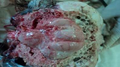
Two cerebral hemispheres are at the anterior part of the African ostrich brain, and the cerebellum and spinal cord are at the posterior part. A cerebral longitudinal fissure is between the 2 cerebral hemispheres and there is a transverse sulcus between the cerebrum and cerebellum. The cerebral surface is smooth, and there is no gyrus or sulcus on it. but, on the dorsal surface of the cerebrum, there is a cambered supersulcus bending posterior-medially, a so-called telencephalic vallecula. The formation of the cerebrum is an obtuse triangle. The cerebellum is located behind the transverse sulcus. An obvious vermis is at the center of the cerebellum with some transverse fissures, and there is a cerebellar auricle on each side of the vermis.

Fig 1. The dorsal view of the African ostrich brain
The ventral view:
in ventral surface of African ostrich brain is an underdeveloped olfactory bulb on each side of the cerebral hemisphere. The optic chiasm occurs at the center back of the 2 cerebral hemispheres. The orbital faces of the cerebral hemisphere are located in the anterior part of the brain. The front portion of the brain stem is the hypothalamus, with a small global hypophysis on it. The midbrain peduncle, the pons, and the medulla oblongata are located consecutively at the back. There is no obvious demarcation between the midbrain and the pons and the medulla oblongata.

Fif 2. The ventral view of the African ostrich brain
The morphological and anatomical characteristics of certain parts of the ostrich brain:
1) Cerebrum: in dorsal view of the brain The cerebrum appears as an obtuse triangle. The surface of the cerebrum is smooth. The posterior part of the cerebral hemisphere is broad, connecting with the optic lobe of the midbrain. The middle of the head of the ostrich’s cerebral hemisphere is a respective cusp, linking to the undeveloped olfactory bulb. The left and right lateral parts of the cerebrum extend laterally, as a hemicycle.
2) Cerebellum: The cerebellum of the African ostrich is developed. the average length of the cerebellum is 34.07 mm, and the average height is 11.70 mm. The length is 2.91 times larger than the height, and The upside of the cerebellum is relatively smooth.
3) Brain stem: The brain stem of the African ostrich, including the midbrain, pons, and medulla oblongata, is similar to those of the other poultries.
Discussion: African ostriches can live in wild deserts with harsh climates and sparse food. The nervous system is the most important synthetic organ system in body of animals. This system plays a prominent role in controlling vital movements of the animal organism. although this is not the only brain function. The duty of cerebellum in body is monitoring and regulating. The shape of the African ostrich cerebellum is mainly different from the other animals. (Romer, 1977). The cerebella of poultries are more developed than those of crawling animals. The length of cerebellum in the African ostrich brain is approximately 2.91 times larger than its height. The posterior superior part of the cerebellar vermis in African ostrich brain approximately forms an angle. Some studies show that injuring the cerebellum will severely impact the accuracy of voluntary movement, including the change of muscular tension, hypomotility, and so on (Ruan et al., 1985). The Table above shows that in African ostriches of weight 90-140 kg, the brain weighs 29.37 g, representing only 0.26% of the body weight. this datum show that the encephalic ratio of the African ostrich is the lowest among the other birds. so we can say The brain of the African ostrich is retarded.

Fig 3. the method of split of the skull skin

Fig 4. cutting lines on the skull

Fig 5. dorsal view of the African ostrich brain in skull
References
1. Saayman, H.S., Naudé, R.J., Oelofsen, W., Isaacson, L.C.: Mesotocin and vasotocin, two neurohypophysial hormones in the ostrich, Struthio camelus. Int. J. Pept. Protein Res., 1986; 28: 398-402.
2. Peng, K.M., Liu, H.Z., Feng, Y.P., He,W.B., Tang,W.H.: African ostrich. Chinese J.Wildlife, 2004; 25: 10. (article in Chinese).
3. Anonymous: Ostrich tastes almost like beef. Misset World Poultry, 1944; 10: 19.
4. Al-Nasser, A., Al-Khalaifa, H., Holleman, K., Al-Ghalaf, W.: Ostrich production in the arid environment of Kuwait. J. Arid Environ., 2003; 54: 219-224.
5. Mushi, E.Z., Isa, J.F., Chabo, R.G., Segaise, T.T.: Growth rate of ostrich (Struthio camelus) chicks under intensive management in Botswana. Trop. Anim. Health Prod., 1998; 30: 197-203.
6. Skadhauge, E., Dawson, A.: The Ostrich Biology, Production and Health. Monks Wood, Abbots Ripton, Huntingdon. CAB Int., 1999: 66.
7. Wang, X.C., Zhang, M.X., Min, H.P.: Histological structure of artificial breeding ostrich skin. China Leather, 2001; 30: 35-38. (article in Chinese).
8. Madekurozwa, M.C.: Morphological features of the luminal surface of the magnum in the sexually immature ostrich (Struthio camelus). Anat. Histol. Embryol., 2005; 34: 350-353.
9. Jin, E.H., Peng, K.M., Wang, J.A, Du, A.N., Tang, L., Wei, L., Wang, Y., Li, S.H., Song, H.: Study on the morphology of the olfactory organ of African ostrich chick. Anat. Histol. Embryol., 2008; 37: 161-165.
10. Peng, K.M., Feng, Y.P., Qiu, D.X.: The comparative anatomy of the skeletons of the ostrich and the domestic fowls. XIV International Congress of Morphological Science. International Academic Publishers. 1997 ; 201.
11. Kiama, S.G., Maina, J.N., Bhattacharjee, J., Mwangi, D.K., Macharia, R.G., Weyrauch, K.D.: Themorphology of the pectin oculi of the ostrich, Struthio camelus. Ann. Anat., 2006; 188: 519-528.
12. Yang, A.F.: Vertebrate Zoology. Pekin University Press. Beijing. 1985; 259-282. (article in Chinese).
13. Liu, Q.E., Li, C.Q., Wang, C.H.:Western and Chinese traditional medicine synthesis treatment study on carpolite accumulating in ostrich stomach. Ostrich Breed. Dev., 1997; 11: 30-32. (article in Chinese).
14. Romer, A.S.: The Vertebrate Body. Philadelphia, Saunders Company, 1977; 516-545.
15. Ruan, D.Y., Shou, T.D.: Neurophysiology. Hefei: China Science and Technology University Press. 1985; 139-159. (article in Chinese).
16. Ma, K.Q., Zheng, G.M.: Comparative Anatomy of Vertebrates. Beijing: Higher Education Press. 1984; 360-400. (article in Chinese).
17. Baumel, J.J., King, A.S., Breazile, J.E., Evans, H.E., Vanden Berge, J.C.: Nomina Anatomica Avium. Cambridge, MA: Nuttall Ornithological Club. 1993; No. 23.
18. Sturkie, P.D.: Poultry Physiology. Science Press. Beijing. 1982; 42-45. (article in Chinese).
19. Yang, X.P.: Animal Physiology. Higher Education Press. Beijing. 2002; 260-312. (article in Chinese).
20. Cong, Z.J., Cong, S.Z.: Synthesis treatment for sand accumulating in stomach of ostrich chicks. Ostrich Breed. Dev., 1998; 12: 22-23. (article in Chinese).
21. Peng, K.M., Niu, H.P., Chen, J.A., Zhang, D.R., Yang, Q.Y., Zhang, J.Y.: Comparative anatomy of the brains of oriental white stork, duck and goose. J. Heilongjiang Agr. Univ., 1994; 7: 81-87. (article in Chinese).
Cite This Work
To export a reference to this article please select a referencing style below:


